How to Incorporate Deep Learning Into Pathology
The medical trade has been exploiting AI for the previous decade or so. We will see a number of approaches to Artificial Intelligence in Pathology that use Whole Slide Images.
In this text, we’ll dissect how to strategy the issues like classification and segmentation for the Whole Slide Images (WSI) that may present insights to pathologists shortly!
Why use AI in Pathology?
The use of digital pathology has turn into very outstanding because the starting of the Information Technology period. Computational AI in pathology can open doorways to many purposes, together with AI. Due to latest advances, it has turn into simpler to develop image-based diagnostic, prognostic, and predictive algorithms.
Some of the primary duties completed by pathologists are classification, segmentation, and quantification. Deep studying strategies elevate these duties dramatically. Apart from gathering extra correct outcomes, investing in AI will assist pathologists be extra productive by lowering human errors, having higher workloads, and faster affected person turnaround time. Additionally, it’s simpler to automate pipelines when coping with the majority of photos.
A short about WSI
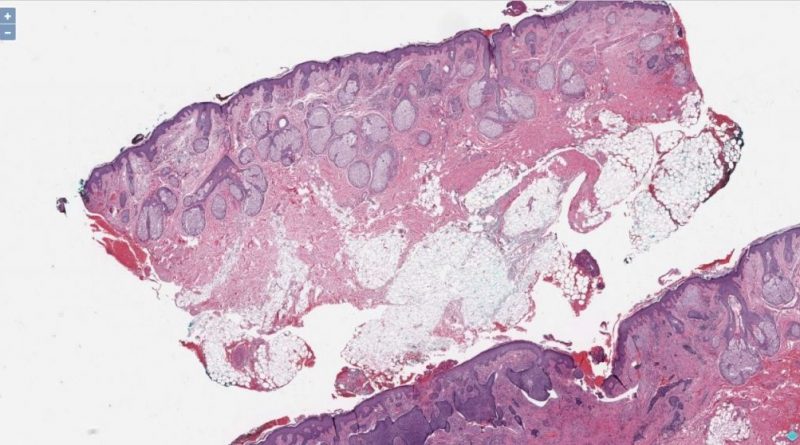
Whole Slide Imaging, also referred to as digital microscopy, goals to imitate microscopy in a computer-generated method. Thus, archiving and making use of image-based options is trouble-free.

The approaches defined beneath are generic and will have utilization in numerous use-cases in pathology. We will look into each the classification and segmentation of WSI.
Normalizing H&E
If you’re working with H&E photos. You might have to observe a further set of processing steps to convert photos from RGB to OD area.
Download the paper right here.
Classification
The WSI decision is often giant, exceeding 15K. It will demand heavy computational energy and reminiscence utilization to prepare a easy classifier on the whole picture.
First Approach
If the classification options are obvious, then resizing and performing vanilla CNN classification will do the trick! It is the best one and takes the least period of time. The issue is that we choose mannequin structure and picture decision primarily based on elements like obtainable compute energy and reminiscence.
Second Approach
It is tiling or patching the Whole Slide Images.
Some libraries on the market that may extract patches of tissues from a WSI similar to CLAM or pathologists’ intervention could also be wanted to annotate the area of curiosity and get these annotations. Therefore, we select the tiles inside this annotated area for coaching and validation.

Classification is utilized to every tile of a specific picture. Ultimately, all predictions will decide the category of the WSI.
The benefit is that tiling produces many photos from a handful of WSI. But it’s extra time-consuming because it requires extra processing efforts to generate tiles.
Models
Preferably, it’s higher to do Transfer Learning due to the complicated options of the pictures. So, fashions like VGG16/ResNet weights are appropriate, or there may very well be open-source pre-trained fashions primarily based on the use case. The switch studying mannequin could converge sooner.
Instead of utilizing pre-trained weights from VGG16/ResNet, we will additionally construct a CNN mannequin from scratch. It is efficient if the picture options are excellent that have to be seen and distinguishable. The mannequin measurement may even be small and will not require heavy useful resource utilization.
Segmentation
The segmentation exhibits the specified components of WSI like tumors, tissues, and wounds that assist pathologists make selections sooner.
Segmentation is the pixel-level classification the place a masks will get generated given a set of lessons. Tiling is critical to carry out segmentation on high-resolution photos similar to WSI.
Generally, the method is as follows,

Tiled strategy for segmentation (do not thoughts the masks)
- With the assistance of annotations, generate a masks for the WSI.
- Run pre-processing (if any) and tile WSIs and their corresponding masks.
- Train the mannequin on the tiled information.
- Run post-processing (if any) after which restitch the generated masks as an entire.
It is critical to set up a normal for WSI decision/dimensions. While coaching a segmentation mannequin, conforming to just one enter form should not be neglected. Therefore, when performing tiling on the unique WSIs, we should guarantee resizing them beforehand.
To make clear, assume that we have now 3 WSIs with completely different resolutions 15640x15640, 15360x16896, and 17920x12000. If the enter form or tile dimension is 512x512, then not all of those are divisible by 512. Then a method could be to resize these WSIs to 15360x15360, which is a a number of of 512.
Albeit, this works nicely when completely different photos’ resolutions are very shut. But when the distinction is important, just like the third decision the place a dimension is 12000, the resized picture will probably be skewed and may lose vital options related for segmentation. Ideally, the WSI decision must be uniform for all WSIs.
Similar to the 2nd strategy of classification, we will select the tiles related to the use case.
Models
There are many segmentation mannequin architectures carried out. But the preferred ones are Unet and Mask-RCNN. These are absolutely able to performing occasion/semantic segmentation over photos.

Unet Model Architecture
Unet was created to carry out segmentation over medical photos. The first benefit of utilizing Unet is that it permits for using world location and context on the similar time. Secondly, it really works with only a few coaching samples and gives higher efficiency for segmentation duties.

However, the one benefit of utilizing Mask-RCNN over Unet could be its simplicity. Unet requires extra processing steps. Overall, there is no such thing as a distinction between Unet and Mask-RCNN outcomes, or which may be particular to the use case.
Regardless of the mannequin structure, segmentation is a resource-demanding activity. Therefore, use a GPU with a strong machine for each coaching and inference.
Hierarchical Approach
Classification and segmentation could be inside a pipeline belonging to completely different steps. Classification may filter out photos that have to be handed to the segmentation mannequin to acquire extra correct outcomes and keep away from redundant processing. Similarly, segmented areas could require additional classification. The pipeline can have many levels like these.

Closure
The goal of this text was to spotlight the points of Deep Learning in pathology. The talked about approaches are generic and might have complicated processes relying on the use circumstances. But they need to give a great baseline to confront your pathology tasks.
The submit How to Incorporate Deep Learning Into Pathology appeared first on Datafloq.

